Educational Disclaimer: This article is for informational and educational purposes only. It does not constitute medical advice, laboratory protocol, or product recommendation. The information presented summarizes published scientific literature on peptide chemistry. Consult a licensed healthcare provider before making any health decisions.
Peptide Stability and Storage: How Degradation Works and How to Prevent It
You can source a peptide with 99% HPLC purity, verified mass spectrometry, clean endotoxin results, and residual solvents well within ICH limits. But if that peptide is stored incorrectly for even a few weeks, none of those quality metrics matter anymore. The compound sitting in the vial is no longer the compound described on the Certificate of Analysis.
Peptide degradation is not hypothetical. It is a well-characterized set of chemical reactions that begin the moment a peptide is exposed to heat, moisture, light, or oxygen. These reactions are predictable, measurable, and in most cases preventable with proper handling.
This guide explains the major degradation pathways that affect research peptides, the critical differences between storing lyophilized powder and reconstituted solutions, the environmental factors that accelerate breakdown, and how to read stability-related data when it appears on a COA.
Why Peptides Are Inherently Unstable
Proteins and peptides are built from amino acid residues linked by amide (peptide) bonds. While the peptide backbone itself is reasonably stable under physiological conditions, the side chains of individual amino acids are not. Many of the 20 standard amino acids contain functional groups that are chemically reactive under conditions that researchers encounter routinely: ambient temperature, dissolved oxygen, neutral pH, and aqueous solution.
This is not a flaw in manufacturing. It is a fundamental property of peptide chemistry. A 15-residue peptide like BPC-157 contains dozens of reactive sites, any one of which can undergo modification under the wrong storage conditions. The same structural flexibility that makes peptides biologically interesting makes them chemically vulnerable.
Understanding the specific degradation pathways helps researchers recognize when a peptide has been compromised, what storage conditions prevent each pathway, and what to look for on analytical reports.
The Four Major Degradation Pathways
Peptide degradation in research settings falls into four primary categories. Each has distinct chemistry, distinct risk factors, and distinct analytical signatures.
1. Deamidation
Deamidation is the single most common chemical degradation pathway for peptides [1]. It occurs when asparagine (Asn) or glutamine (Gln) residues lose their amide group, converting to aspartic acid (Asp) or glutamic acid (Glu), respectively.
The chemistry: The asparagine side chain undergoes a spontaneous intramolecular rearrangement through a cyclic succinimide intermediate. This intermediate can then hydrolyze to form either aspartate or isoaspartate, the latter being a non-standard amino acid linkage that can significantly alter peptide conformation and biological activity [1][2].
What accelerates it:
- Elevated pH (above 6.0). The reaction is base-catalyzed.
- Higher temperatures. The rate roughly doubles for every 10°C increase.
- Aqueous solution. Water is a direct reactant in the hydrolysis step.
- Sequence context. Asn-Gly sequences are particularly susceptible because the small glycine side chain offers minimal steric hindrance to succinimide formation [2].
Why it matters: Deamidation introduces a negative charge where there was none (asparagine is neutral; aspartate carries a -1 charge at physiological pH). This charge change can disrupt electrostatic interactions critical to the peptide's three-dimensional structure. For peptides studied in receptor-binding assays or cell culture, even a single deamidation event can measurably alter activity [1].
How it appears on analytics: Deamidation adds approximately 1 Da of mass per event (the amide NH2 is replaced by OH). On HPLC, deamidated species typically elute as a shoulder or secondary peak near the parent compound, because the charge change slightly alters retention time. Mass spectrometry will show a +0.984 Da shift from the theoretical mass [3].
2. Oxidation
Oxidation is the second most prevalent degradation pathway and the most common cause of peptide degradation during storage in solution [4].
The chemistry: Methionine (Met) residues are the primary target. The thioether sulfur in methionine is oxidized to methionine sulfoxide (Met(O)) by dissolved oxygen, peroxides, or trace metal ions acting as catalysts. Tryptophan (Trp) and cysteine (Cys) residues are also susceptible, with tryptophan oxidizing to various kynurenine and hydroxytryptophan derivatives, and free cysteine forming disulfide bonds or sulfenic/sulfinic acid species [4][5].
What accelerates it:
- Dissolved oxygen. Even degassed solutions re-equilibrate with atmospheric oxygen within hours unless sealed under inert gas.
- Light exposure. UV light (particularly 254-280 nm) generates reactive oxygen species that dramatically accelerate Met and Trp oxidation.
- Trace metals. Iron, copper, and other transition metals catalyze Fenton-type reactions that produce hydroxyl radicals. These metals can leach from glass vials or contaminate bacteriostatic water.
- Higher temperatures. As with most chemical reactions, oxidation rates increase with temperature.
- Alkaline pH. Oxidation is generally faster above pH 7.
Why it matters: Methionine oxidation is particularly damaging because it introduces a bulky, polar sulfoxide group that can disrupt hydrophobic packing within the peptide structure. For peptides where methionine participates in receptor binding or is located near the active site, oxidation can reduce biological activity by 50-90% [4]. Tryptophan oxidation is often accompanied by visible changes: the solution may develop a yellow or brown tint.
How it appears on analytics: Each Met oxidation adds +16 Da (addition of one oxygen atom). HPLC typically shows an earlier-eluting peak because the sulfoxide is more hydrophilic than the parent thioether. Mass spectrometry is definitive: the +16 Da mass shift is unambiguous [3][5].
3. Hydrolysis
Hydrolysis is the cleavage of the peptide bond itself, breaking the chain into two or more fragments.
The chemistry: The amide bond connecting adjacent amino acids is thermodynamically unstable in aqueous solution, though kinetically slow under mild conditions. Certain sequences are far more labile. Asp-Pro bonds are notoriously susceptible to acid-catalyzed cleavage, and bonds adjacent to aspartate residues are vulnerable across a wide pH range due to the same succinimide intermediate involved in deamidation [6].
What accelerates it:
- Extreme pH. Both strongly acidic (below pH 2) and strongly alkaline (above pH 10) conditions accelerate backbone hydrolysis.
- High temperatures. Autoclaving or prolonged heating above 40°C can cause detectable cleavage.
- Extended time in solution. Even at neutral pH and refrigerated temperatures, hydrolysis accumulates over weeks to months.
Why it matters: Unlike deamidation or oxidation, which modify a peptide, hydrolysis destroys it. The fragments may retain partial activity, no activity, or unexpected activity depending on which bond was cleaved. Hydrolysis is irreversible, and there is no way to restore a cleaved peptide bond outside of re-synthesis.
How it appears on analytics: Hydrolysis produces new peaks on HPLC at different retention times from the parent. Mass spectrometry shows two (or more) species whose masses sum to the parent mass plus 18 Da (the water molecule consumed in cleavage). A sudden drop in HPLC purity from, say, 98% to 85% with new low-molecular-weight peaks is a strong indicator of hydrolytic degradation [6].
4. Aggregation
Aggregation is the self-association of peptide molecules into dimers, oligomers, or insoluble particulates.
The chemistry: Aggregation can proceed through covalent or non-covalent mechanisms. Covalent aggregation most commonly involves disulfide bond formation between cysteine residues on separate peptide molecules. Non-covalent aggregation is driven by hydrophobic interactions, where partially unfolded peptides expose hydrophobic surfaces that associate in aqueous solution [7].
What accelerates it:
- High peptide concentration. The higher the concentration, the more frequent molecular collisions become.
- Elevated temperature. Heat promotes partial unfolding, exposing aggregation-prone regions.
- Agitation. Mechanical stress (shaking, vortexing, repeated freeze-thaw cycles) can denature peptides at the air-water interface, promoting aggregation.
- pH near the isoelectric point. At their pI, peptides carry no net charge and electrostatic repulsion between molecules is minimized.
Why it matters: Aggregated peptide is functionally lost. Dimers and oligomers may not bind receptors with the same affinity as monomers. Insoluble aggregates are completely inactive and may also trigger immune responses in biological assays, introducing confounding variables [7].
How it appears on analytics: Size-exclusion chromatography (SEC) is the primary method for detecting aggregation, showing peaks at higher molecular weight than the monomer. On standard RP-HPLC, aggregates may appear as broad, poorly resolved peaks or may not elute at all (remaining stuck on the column). Visual inspection can detect advanced aggregation: turbidity, visible particles, or gel formation in a previously clear solution.
Lyophilized vs. Reconstituted: Two Very Different Stability Profiles
The single most important variable in peptide storage is whether the compound is in its dry (lyophilized) state or dissolved in solution. The stability difference between these two states is not incremental. It is typically measured in orders of magnitude.
Lyophilized (Dry Powder) Stability
Lyophilized peptides are produced by freeze-drying purified peptide solutions. The process removes water, leaving a dry cake or powder that is far more resistant to degradation than the corresponding solution.
Why lyophilized peptides are more stable:
- Water is removed. Deamidation and hydrolysis both require water as a reactant. In the absence of free water, these reactions are suppressed by orders of magnitude [8].
- Molecular mobility is restricted. In the solid state, peptide molecules are essentially frozen in place. The molecular collisions required for aggregation cannot occur. Intramolecular rearrangements (like succinimide formation) are dramatically slowed.
- Oxidation still occurs but at reduced rates, because dissolved oxygen is not present. Residual oxygen in the headspace of a sealed vial can still cause slow surface oxidation, which is why quality suppliers seal vials under nitrogen or argon [8].
Typical shelf life: Properly lyophilized and sealed peptides stored at -20°C are generally stable for 2-5 years with minimal degradation. At 4°C (refrigerator), most lyophilized peptides remain stable for 12-24 months. At room temperature, stability varies widely by sequence but is typically measured in months rather than years [8][9].
Key risk factors for lyophilized peptides:
- Moisture ingress. The lyophilized cake is hygroscopic. If the seal is compromised or the vial is repeatedly opened in a humid environment, the cake absorbs atmospheric moisture and degradation pathways reactivate.
- Temperature cycling. Repeated freeze-thaw of the sealed vial is not harmful to dry powder, but the temperature fluctuations can cause condensation inside the vial if the seal is imperfect.
- Residual moisture content. Not all lyophilization cycles are equally effective. A well-optimized cycle produces a cake with less than 1-2% residual moisture. A poorly controlled cycle may leave 5-10%, significantly reducing stability. Residual moisture content is sometimes reported on COAs and is always worth checking [8].
Reconstituted (In-Solution) Stability
Once a lyophilized peptide is reconstituted in bacteriostatic water, sterile water, or buffer, all four degradation pathways become active. The clock starts immediately.
Typical stability windows for reconstituted peptides:
- Refrigerated (2-8°C): Most reconstituted peptides remain analytically stable for 2-4 weeks. Some particularly stable sequences may last 6-8 weeks; susceptible sequences (those with Asn-Gly motifs, multiple Met residues, or free Cys) may degrade measurably within days [9][10].
- Room temperature (20-25°C): Hours to days for sensitive peptides; 1-2 weeks maximum for robust sequences. Not recommended for storage.
- Frozen (-20°C): Reconstituted peptide aliquots can be frozen to extend stability to several months. However, repeated freeze-thaw cycles are damaging (see below).
Why reconstituted peptides degrade faster:
- Water is present, enabling deamidation and hydrolysis.
- Dissolved oxygen is present, enabling oxidation.
- Molecular mobility is restored, enabling aggregation.
- pH effects are active. The reconstitution solvent's pH directly influences degradation rates.
The freeze-thaw problem: Freezing a reconstituted peptide solution is an effective strategy for extending shelf life, but each freeze-thaw cycle introduces stress. During freezing, ice crystal formation concentrates the peptide and any salts or buffers into an increasingly small liquid phase (cryoconcentration). This can cause local pH shifts, dramatically elevated peptide concentration, and ice-liquid interface stress that promotes aggregation and denaturation [10].
Best practice: Aliquot reconstituted peptide into single-use volumes before freezing. This eliminates the need for repeated freeze-thaw cycles entirely. Use low-binding microcentrifuge tubes or polypropylene vials. Label each aliquot with the peptide identity, concentration, reconstitution date, and solvent used.
Environmental Factors: A Practical Reference
Temperature
Temperature is the most controllable and most impactful storage variable.
Storage Condition | Lyophilized Stability | Reconstituted Stability
-80°C (ultra-low freezer) | 5+ years | 6-12 months (single freeze)
-20°C (standard freezer) | 2-5 years | 3-6 months (single freeze)
2-8°C (refrigerator) | 12-24 months | 2-4 weeks (typical)
20-25°C (room temperature) | 3-12 months (sequence-dependent) | Days to 1-2 weeks
37°C (incubator/shipping stress) | Weeks to months | Hours to days
These ranges are generalizations. Actual stability depends on the specific peptide sequence, purity, formulation, and container closure system. They represent the ranges most commonly reported in pharmaceutical stability literature for peptides in the 1-5 kDa molecular weight range [8][9][10].
Light
Ultraviolet and visible light drive photodegradation, primarily targeting tryptophan and tyrosine residues. Tryptophan absorbs strongly at 280 nm and is vulnerable to photooxidation that produces kynurenine and other degradation products. Tyrosine can undergo photo-induced cross-linking.
Practical guidance: Store peptide vials in opaque containers or wrapped in aluminum foil. If the original packaging is amber glass, leave it in the vial. Do not store reconstituted peptides in clear glass or plastic containers on open benchtops under fluorescent lighting. Even indirect sunlight through a laboratory window can accelerate degradation of Trp-containing peptides measurably within days [5].
Oxygen
Atmospheric oxygen (21% O2) is the primary driver of methionine and tryptophan oxidation. Dissolved oxygen in reconstitution solvents adds further risk.
Practical guidance: Quality suppliers seal lyophilized vials under inert gas (nitrogen or argon). When reconstituting, minimize the headspace in the vial. If you are aliquoting into multiple tubes, fill them as completely as practical and consider overlaying with nitrogen using a gentle stream from a gas cylinder. For high-value or oxidation-sensitive peptides, oxygen-free reconstitution using degassed, nitrogen-sparged solvents is worth the effort [4][5].
pH
The reconstitution solvent's pH influences nearly every degradation pathway.
- Deamidation accelerates above pH 6 and is fastest at pH 7-8.
- Oxidation is generally faster at alkaline pH.
- Hydrolysis of Asp-X bonds accelerates below pH 4; backbone hydrolysis accelerates at pH extremes.
- Aggregation is often maximal near the peptide's isoelectric point.
Practical guidance: Most research peptides are reconstituted in bacteriostatic water (pH ~5-7) or sterile water (pH ~5.5-7.0). These slightly acidic to neutral conditions represent a reasonable compromise for most sequences. For peptides known to be deamidation-sensitive (those containing Asn-Gly or Asn-Ser motifs), reconstitution in mildly acidic buffer (pH 4-5) can significantly extend solution stability [1][2].
Container Material
The vial or tube material matters more than most researchers realize.
- Glass: Borosilicate glass is standard for lyophilized peptide vials. It is chemically inert, provides a good oxygen barrier, and is compatible with most peptides. However, glass surfaces can leach trace metals (particularly from soda-lime glass) and can adsorb peptides at low concentrations, reducing effective yield.
- Polypropylene: Low-binding polypropylene tubes are preferred for reconstituted aliquots. They minimize surface adsorption compared to polystyrene or standard polypropylene. At peptide concentrations below 0.1 mg/mL, surface adsorption losses can exceed 20-30% in standard tubes [11].
- Avoid polystyrene. Polystyrene tubes have high peptide binding and are not suitable for peptide storage.
How to Interpret Stability Data on a COA
Not all COAs include stability data, but when they do, it provides valuable information about the peptide's degradation profile and the supplier's quality practices.
What to Look For
Purity at release vs. purity at retest: Some COAs report HPLC purity at the time of manufacture ("release") and again at a later retest date. Comparing these two numbers gives you a direct measure of degradation over time. A peptide that was 99.1% pure at release and 98.7% at a 6-month retest under recommended storage conditions shows minimal, acceptable degradation. A drop from 99% to 92% in the same timeframe signals a stability problem.
Related substances or impurity profiles: Detailed COAs may list specific impurity peaks as a percentage of total area. Tracking whether these impurities increase between release and retest tells you which degradation pathways are active. A growing peak at +16 Da (detected by LC-MS) points to oxidation. A growing peak at +1 Da points to deamidation.
Storage conditions stated on the COA: The COA should specify under what conditions the peptide was tested and under what conditions it should be stored. If the COA states "Store at -20°C, protect from light" and you have been keeping the vial at 4°C on an open shelf, the analytical data on the COA no longer represents what is in your vial.
Moisture content: Some COAs report Karl Fischer titration results for residual moisture in the lyophilized powder. Values below 2% are typical for well-lyophilized peptides. Values above 5% suggest the lyophilization cycle was suboptimal and the peptide will have reduced shelf life compared to a properly dried product [8].
Appearance: The physical description of the lyophilized cake can indicate stability. A fluffy white or off-white cake is normal. A collapsed, glassy, or discolored cake may indicate exposure to moisture or heat during storage or shipping. Yellow discoloration in a Trp-containing peptide specifically suggests photo-oxidation.
What the COA Cannot Tell You
A COA is a snapshot at a moment in time. It does not guarantee the peptide's condition at the time you receive and open it. Shipping conditions, warehouse temperature control, time in transit, and handling between the COA date and your receipt all introduce variables that the COA cannot account for.
This is why storage conditions after receipt matter so much. The COA tells you what the peptide was when it left the supplier. Maintaining appropriate storage conditions preserves that quality. Improper storage degrades it, regardless of what the COA says.
Practical Storage Protocol for Research Peptides
Based on the degradation chemistry described above, here is a consolidated set of storage practices that apply to the majority of research peptides in the 1-5 kDa range.
For Unopened Lyophilized Vials
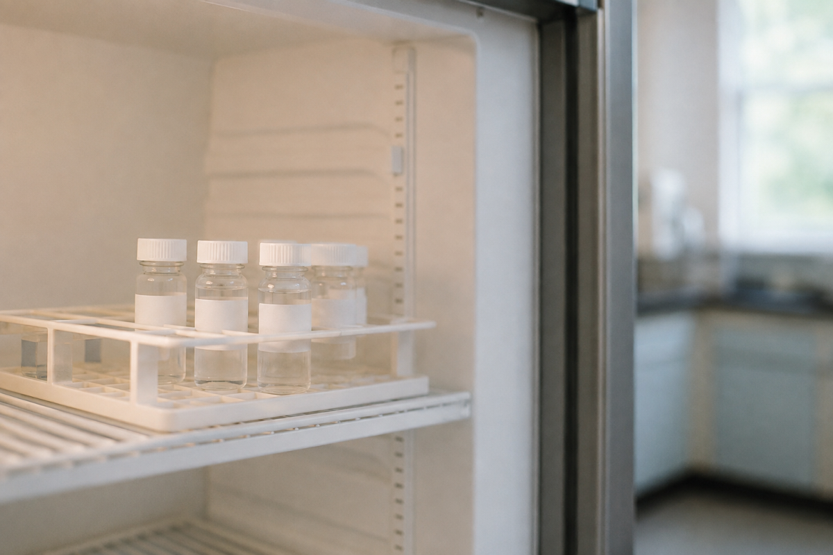
- Store at -20°C or colder. Ultra-low (-80°C) is ideal for long-term storage but not required for most peptides kept under 2 years.
- Keep the original seal intact. The crimped cap and butyl rubber stopper provide a moisture and oxygen barrier. Do not remove the cap until you are ready to reconstitute.
- Protect from light. Store in the original box or wrap in foil if the vial is clear glass.
- Allow the vial to reach room temperature before opening. Removing a cold vial from the freezer and immediately puncturing the septum creates a pressure differential that draws humid air into the vial, potentially introducing moisture to the lyophilized cake.
For Reconstituted Peptides
- Reconstitute with appropriate solvent. Bacteriostatic water (0.9% benzyl alcohol) is standard for peptides that will be used over multiple days. Sterile water for injection is appropriate for single-use reconstitution.
- Aliquot immediately. Divide the reconstituted solution into single-use volumes in low-binding polypropylene tubes. Label with peptide name, concentration, date, and solvent.
- Store aliquots at -20°C. Thaw one aliquot per use. Do not refreeze thawed aliquots.
- If refrigerating for short-term use (days, not weeks): Keep at 2-8°C, protect from light, and use within 2-4 weeks. Monitor for turbidity, color change, or particulate formation.
- Minimize headspace. Use appropriately sized containers. Excess air in the container means more dissolved oxygen.
- Never store reconstituted peptides at room temperature unless actively using them within hours.
Reconstitution Math: A Quick Reference
When reconstituting a lyophilized peptide, the concentration you choose affects both usability and stability. Higher concentrations are generally more stable (less surface adsorption loss per microgram) but may exceed the peptide's solubility. Lower concentrations are more susceptible to adsorption losses and may degrade faster.
A common starting point: add bacteriostatic water to achieve a concentration of 1-5 mg/mL, depending on the peptide's solubility. Most research peptides in the 1-5 kDa range are freely soluble at these concentrations in aqueous solvents. Consult the product datasheet or COA for peptide-specific solubility data.
Sequence-Specific Vulnerability: Which Peptides Need Extra Care?
Not all peptides degrade at the same rate. Sequence composition determines which degradation pathways are active and how fast they proceed.
High deamidation risk: Peptides containing Asn-Gly, Asn-Ser, Asn-Ala, or Asn-His sequences. The smaller the amino acid following asparagine, the faster the deamidation. Glutamine residues deamidate more slowly than asparagine but are not immune [1][2].
High oxidation risk: Peptides containing methionine, tryptophan, or free cysteine (not disulfide-bonded). Met-containing peptides are the most common concern in research catalogs. Trp-containing peptides are additionally photosensitive [4][5].
High hydrolysis risk: Peptides containing Asp-Pro bonds. This specific dipeptide motif is unusually labile under mildly acidic conditions. Peptides with multiple aspartate residues adjacent to small amino acids are also at elevated risk [6].
High aggregation risk: Hydrophobic peptides, cysteine-containing peptides (disulfide-mediated aggregation), and peptides with high beta-sheet propensity. Longer peptides (>30 residues) are generally more aggregation-prone than shorter ones [7].
For researchers working with specific compounds from the PeptidesEuro catalog: checking the amino acid sequence against these risk factors helps predict which degradation pathways to monitor and which storage conditions to prioritize. This information, combined with the analytical methods described in our HPLC and mass spectrometry guides, provides a complete framework for assessing and maintaining peptide quality over time.
Signs That a Peptide Has Degraded
Researchers should watch for these indicators that a stored peptide may no longer match its original quality profile:
- Visual changes in lyophilized powder: Discoloration (yellow, brown), collapsed cake structure, visible moisture or droplets inside the vial, crystalline appearance replacing the original fluffy cake.
- Visual changes in solution: Turbidity, visible particles, gel formation, color change (particularly yellowing in Trp-containing peptides), precipitation.
- Analytical changes: Drop in HPLC purity percentage. Appearance of new impurity peaks. Mass shift detected by LC-MS. Broadened or shifted peaks on RP-HPLC.
- Functional changes: Reduced activity in bioassays. Inconsistent results between fresh and stored preparations. Unexpected cytotoxicity (which can indicate aggregation).
If any of these signs are present, the peptide should be re-analyzed before use in critical experiments, or replaced with a freshly reconstituted aliquot from a properly stored lyophilized source.
The Bottom Line
Peptide stability is not a mystery. It is governed by well-understood chemical reactions that are predictable, measurable, and preventable. The four major degradation pathways, deamidation, oxidation, hydrolysis, and aggregation, each respond to specific environmental triggers. Removing those triggers through appropriate storage is the single most effective way to protect the investment in a quality peptide.
Lyophilized peptides stored sealed at -20°C, protected from light and moisture, represent the most stable form. Once reconstituted, the clock starts: aliquot immediately, freeze what you won't use within days, and discard anything that shows signs of degradation.
The COA tells you where the peptide started. Storage determines where it ends up.
References
[1] Capasso, S., et al. "Deamidation of Asparagine and Glutamine Residues in Peptides and Proteins." Journal of Peptide Science, vol. 6, no. 5, 2000, pp. 227-235.
[2] Robinson, N.E., Robinson, A.B. "Molecular Clocks: Deamidation of Asparaginyl and Glutaminyl Residues in Peptides and Proteins." Althouse Press, 2004.
[3] Manning, M.C., et al. "Stability of Protein Pharmaceuticals: An Update." Pharmaceutical Research, vol. 27, 2010, pp. 544-575.
[4] Li, S., et al. "Chemical Instability of Protein Pharmaceuticals: Mechanisms of Oxidation and Strategies for Stabilization." Biotechnology and Bioengineering, vol. 48, no. 5, 1995, pp. 490-500.
[5] Kerwin, B.A., Remmele, R.L. "Protect from Light: Photodegradation and Protein Biologics." Journal of Pharmaceutical Sciences, vol. 96, no. 6, 2007, pp. 1468-1479.
[6] Landon, M. "Cleavage at Aspartyl-Prolyl Bonds." Methods in Enzymology, vol. 47, 1977, pp. 145-149.
[7] Wang, W. "Protein Aggregation and Its Inhibition in Biopharmaceutics." International Journal of Pharmaceutics, vol. 289, no. 1-2, 2005, pp. 1-30.
[8] Carpenter, J.F., et al. "Rational Design of Stable Lyophilized Protein Formulations: Some Practical Advice." Pharmaceutical Research, vol. 14, no. 8, 1997, pp. 969-975.
[9] Chi, E.Y., et al. "Physical Stability of Proteins in Aqueous Solution: Mechanism and Driving Forces in Nonnative Protein Aggregation." Pharmaceutical Research, vol. 20, no. 9, 2003, pp. 1325-1336.
[10] Bhatnagar, B.S., et al. "Protein Stability During Freezing: Separation of Stresses and Mechanisms of Protein Stabilization." Pharmaceutical Development and Technology, vol. 12, no. 5, 2007, pp. 505-523.
[11] Goebel-Stengel, M., et al. "The Importance of Using the Correct Syringe Volume in Peptide Research." Analytical Biochemistry, vol. 414, no. 2, 2011, pp. 312-314.
This article is part of PeptidesEuro's Quality & Testing Education series. For related content, see: How to Read a COA for Research Peptides, HPLC vs Mass Spectrometry for Peptide Analysis, Why Residual Solvent Testing Matters, and What Endotoxin Testing Means for Quality.